Foot Muscles Mri - When The Presence Of A Flexor Hallucis Accessory Longus Contributes To Chronic Foot Pain With Standing Podiatry Today. Dorsal interossei (foot) dr yuranga weerakkody ◉ and dr geon oh et al. The gold standard in diagnostic imaging of muscle injuries is magnetic resonance imaging (mri). The adductor hallucis has two heads: Indications for foot mri scan. Adductor hallucis is anatomically located in the central compartment of foot, but the muscle is functionally grouped with the medial plantar muscles of foot because it acts on the great toe (hallux).
Shoulder elbow wrist finger thumb. They are named extensor digitorum brevis and extensor hallucis brevis. Your doctor, with the help of a radiologist, can then examine these images to determine whether there is anything wrong with your foot or ankle. A case report and review of anatomy. Dorsal interossei (foot) dr yuranga weerakkody ◉ and dr geon oh et al.
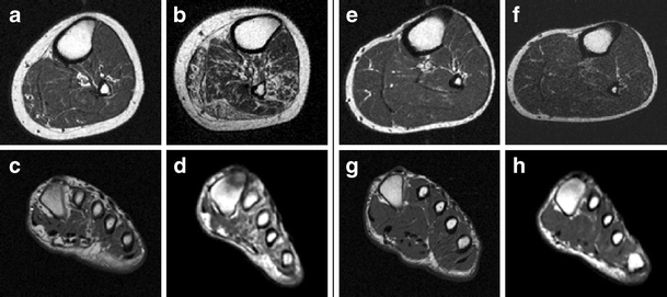
Figure 1 Accelerated Atrophy Of Lower Leg And Foot Muscles A Follow Up Study Of Long Term Diabetic Polyneuropathy Using Magnetic Resonance Imaging Mri Springerlink from media.springernature.com A case report and review of anatomy. Adductor hallucis is anatomically located in the central compartment of foot, but the muscle is functionally grouped with the medial plantar muscles of foot because it acts on the great toe (hallux). Those fibers of the most medial and largest belly are… The interosseous muscles of the foot are muscles found near the metatarsal bones that help to control the toes. The most common ossicle is the os trigonum, which is a prominent unfused apophysis of the lateral tubercle of the talus. In addition, an image of all the muscles of the back and plantar part of the foot, all tendons and tendon ligaments, blood vessels and nerves are obtained. Indications for foot mri scan. They are named extensor digitorum brevis and extensor hallucis brevis.
Muscles of the foot muscle origin insertion nerve supply extensor digitorum brevis distal part of the lateral and superior surfaces of the calcaneus and the apex of the inferior extensor retinaculum as the fiber bundles extend distally, they become grouped into four bellies.
The majority of soft tissue lesions in the foot and ankle are benign. Both muscles are innervated by the deep fibular nerve. Your doctor, with the help of a radiologist, can then examine these images to determine whether there is anything wrong with your foot or ankle. The three plantar interossei muscles adduct the 3 rd, 4 th and 5 th toes toward the long axis through the 2 nd toe. Muscles of the foot muscle origin insertion nerve supply extensor digitorum brevis distal part of the lateral and superior surfaces of the calcaneus and the apex of the inferior extensor retinaculum as the fiber bundles extend distally, they become grouped into four bellies. Shoulder elbow wrist finger thumb. Muscle damage may cause muscle pain and muscle weakness may cause difficulty lifting the arms above the shoulders, climbing stairs, or arising from a sitting position. The four dorsal interossei muscles are the most superior muscles in the sole of the foot and abduct the 2 nd to 4 th toes relative to the long axis through the second toe. The gold standard in diagnostic imaging of muscle injuries is magnetic resonance imaging (mri). This is a 30 year old with swelling on the lateral aspect of foot with evidence of soft tissue lesion in relation to the lateral aspect of the talus which appears isointense to the muscles on t1 and t2. Mri is the choice of modality for further imaging the ankle and foot after obtaining initial radiographs. With a muscle injury, for example, mri images often show a bright signal indicating that there is more water in the muscle, which is a sign of injury. They are mainly responsible for assisting some of the extrinsic muscles in their actions.
In the foot and ankle many accessory ossicles can be seen. This imaging technique assesses the ligaments and tendons, neurovascular structures (tarsal tunnel and plantar fascia), and the osseous structures(19). They are mainly responsible for assisting some of the extrinsic muscles in their actions. Findings on conventional arthrography and mr imaging. Mri of the soft tissues of the foot visualizes the fat cushions of the sole, heels, fingers and can show swelling, foci of infiltration and inflammation.
Baxter S Nerve Entrapment Diagnosis Treatment Injection Surgery from www.fasciitis.com Magnetic resonance imaging (mri) is the modality of choice in diagnosing accessory muscles, delineating their relationship to adjacent structures, and differentiating them from soft tissue tumors. The adductor hallucis has two heads: They are mainly responsible for assisting some of the extrinsic muscles in their actions. In the foot and ankle many accessory ossicles can be seen. Muscle injuries of the hip and thigh are a highly relevant issue in competitive sports imaging. Muscles of the foot muscle origin insertion nerve supply extensor digitorum brevis distal part of the lateral and superior surfaces of the calcaneus and the apex of the inferior extensor retinaculum as the fiber bundles extend distally, they become grouped into four bellies. They are named extensor digitorum brevis and extensor hallucis brevis. Radiologists need to be familiar with typical mri findings in order to accurately detect and classify muscle injuries.
Mri of the ankle and feet
The aim of this review is to provide the reader with a comprehensive overview of the magnetic resonance imaging (mri) characteristics of the most common benign and malignant soft tissue neoplasms which occur around the foot and ankle. Muscle damage may cause muscle pain and muscle weakness may cause difficulty lifting the arms above the shoulders, climbing stairs, or arising from a sitting position. Mri is the choice of modality for further imaging the ankle and foot after obtaining initial radiographs. Mri is an ideal method for identifying areas of muscle atrophy and fatty infiltration. • muscle edema is seen secondary to multiple etiologies including trauma, infectious and inflammatory processes, autoimmune disorders, neoplasms, and denervation injuries • on mri muscle edema is characterized by increase in free water within the muscle • muscle edema is seen on mri as increased signal on fluid sensitive sequences t2 fs Mri of the soft tissues of the foot visualizes the fat cushions of the sole, heels, fingers and can show swelling, foci of infiltration and inflammation. Indications for foot mri scan. Accessory muscles are isointense to skeletal muscle on all pulse sequences, and can insert by fleshy muscular or tendinous insertions. The most common ossicle is the os trigonum, which is a prominent unfused apophysis of the lateral tubercle of the talus. They are mainly responsible for assisting some of the extrinsic muscles in their actions. In addition, an image of all the muscles of the back and plantar part of the foot, all tendons and tendon ligaments, blood vessels and nerves are obtained. Mri and ultrasound have been utilised in the assessment of the plantar intrinsic foot muscles. The adductor hallucis has two heads:
This is a 30 year old with swelling on the lateral aspect of foot with evidence of soft tissue lesion in relation to the lateral aspect of the talus which appears isointense to the muscles on t1 and t2. Those fibers of the most medial and largest belly are… Mri of the ankle and feet The gold standard in diagnostic imaging of muscle injuries is magnetic resonance imaging (mri). The interosseous muscles of the foot are muscles found near the metatarsal bones that help to control the toes.
Role Of Intrinsic Muscle Atrophy In The Etiology Of Claw Toe Deformity In Diabetic Neuropathy May Not Be As Straightforward As Widely Believed Diabetes Care from care.diabetesjournals.org Dorsal interossei (foot) dr yuranga weerakkody ◉ and dr geon oh et al. In addition, an image of all the muscles of the back and plantar part of the foot, all tendons and tendon ligaments, blood vessels and nerves are obtained. The most common ossicle is the os trigonum, which is a prominent unfused apophysis of the lateral tubercle of the talus. Mri is an ideal method for identifying areas of muscle atrophy and fatty infiltration. Mri of the ankle and feet In the foot and ankle many accessory ossicles can be seen. Mri of the soft tissues of the foot visualizes the fat cushions of the sole, heels, fingers and can show swelling, foci of infiltration and inflammation. Muscle anatomy basics 12 photos of the muscle anatomy basics basics of muscle anatomy, muscle anatomy basics, human muscles, basics of muscle anatomy, muscle anatomy basics
A magnetic resonance imaging (mri) was performed on a normal subject;
Magnetic resonance imaging, otherwise known as mri, uses a combination of magnetic fields and radio waves to take images of the internal structures of your body. Those fibers of the most medial and largest belly are… The intrinsic muscles of the foot are key contributors to foot function and are important to evaluate in lower limb disorders. Mri of the ankle and feet The muscles of the dorsum of the foot are a group of two muscles, which together represent the dorsal foot musculature. 9 yao l, do hm, cracchiolo a, et al. Anatomical structures of the ankle and foot and specific regions (major joints) are visible as dynamic labeled images. Shoulder elbow wrist finger thumb. Routine ankle magnetic resonance imaging (mri) tests involve taking images of the foot and ankle in the axial, coronal, and sagittal planes parallel to the tabletop(2). A magnetic resonance imaging (mri) was performed on a normal subject; Magnetic resonance imaging (mri) is the modality of choice in diagnosing accessory muscles, delineating their relationship to adjacent structures, and differentiating them from soft tissue tumors. The interosseous muscles of the foot are muscles found near the metatarsal bones that help to control the toes. With a muscle injury, for example, mri images often show a bright signal indicating that there is more water in the muscle, which is a sign of injury.
Share :
Post a Comment
for "Foot Muscles Mri - When The Presence Of A Flexor Hallucis Accessory Longus Contributes To Chronic Foot Pain With Standing Podiatry Today"


Post a Comment for "Foot Muscles Mri - When The Presence Of A Flexor Hallucis Accessory Longus Contributes To Chronic Foot Pain With Standing Podiatry Today"