Hip Muscles Diagram : Anatomy Hip Joint Muscles - ProProfs Quiz. There are anterior muscles diagrams and posterior muscles diagrams. Hip muscles diagram, learn more about hip muscles diagram. Depending on the situation, with the pelvis in a fixed position the muscles move around the thigh. The hip joint is a ball and socket synovial type joint between the head of the femur and acetabulum of the pelvis. Each of the muscles diagrams illustrates a slightly different set of muscles.
*click them to make them larger & view details. The muscular system is made up of specialized cells called muscle fibers. Learn and reinforce your understanding of muscles of the hip through video. Press into the feet, lengthening the legs to press the hips up toward the ceiling. Bones in human anatomy, the hip flexors are a group of skeletal muscles that act to flex the femur (thigh bone) hip muscles diagram.
Muscular System Quizzes • Anatomy & Physiology from www.getbodysmart.com Press into the feet, lengthening the legs to press the hips up toward the ceiling. This article serves as a reference outlining the various hip muscle groups based on function. The hips are the central pivot point of the entire body, supporting its weight during movement and when standing. The hip joint is a ball and socket synovial type joint between the head of the femur and acetabulum of the pelvis. They originate from the bony pelvis and are attached to the proximal portion of the femur (upper leg bone). Muscles, connected to bones or muscles that act on the lower limb cause movement at the hip, knee and foot joints. Comprehensive information about hip joint anatomy including muscles, tendons, ligaments, bones, bursae, skeletal structure and joint capsules. *click them to make them larger & view details.
A hip flexor and mild hip lateral rotator.
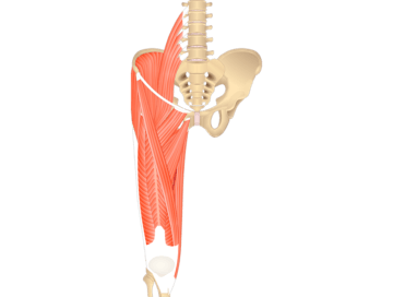
The main muscles of the hip and pelvis consistsof the iliopsoas, pectinues, rectus femoris and sartorius at the front. *click them to make them larger & view details. Diagram representing the anterior view of the muscle groups adductor brevis, adductor longus and adductor magnus. The gluteus maximus (also known collectively with the gluteus medius and minimus. Review muscle diagram using the 2 diagrams below: Flexors, extensors, adductors, abductors, lateral rotators. Comprehensive information about hip joint anatomy including muscles, tendons, ligaments, bones, bursae, skeletal structure and joint capsules. Now that you watched the video, you. If you are starting to feel hip pain or stiffness, you'll want to know hip anatomy: In human anatomy, the muscles of the hip joint are those muscles that cause movement in the hip. The following diagram illustrates the actions of the terms adduction, abduction, flexion and extension at the different joints. Learn vocabulary, terms and more with flashcards, games and other study tools. Muscles, connected to bones or muscles that act on the lower limb cause movement at the hip, knee and foot joints.
Learn vocabulary, terms and more with flashcards, games and other study tools. This is the largest of the three compartments of the thigh. Depending on the situation, with the pelvis in a fixed position the muscles move around the thigh. The gluteus medius, gluteus minimus, piriformis, tensor fasciae latae on the outside. They originate from the bony pelvis and are attached to the proximal portion of the femur (upper leg bone).
A schematic diagram of the posterior view of the left hip shows the... | Download Scientific Diagram from www.researchgate.net The muscles of the hip and thigh keep your hip joints strong and mighty, allowing for a wide range of hip movements. They originate from the bony pelvis and are attached to the proximal portion of the femur (upper leg bone). They are among the largest and strongest muscle in the body and are. Each of these muscles plays a role in the movement or stability of the hip. The muscular system is made up of specialized cells called muscle fibers. Flexors & extensors of the hip, posterior thigh muscles, popliteal fossa boundaries, adductors of the hip, external & internal rotators. Muscles, connected to bones or muscles that act on the lower limb cause movement at the hip, knee and foot joints. The gluteus medius, gluteus minimus, piriformis, tensor fasciae latae on the outside.
Comprehensive information about hip joint anatomy including muscles, tendons, ligaments, bones, bursae, skeletal structure and joint capsules.
Smartdraw includes 1000s of professional healthcare and anatomy chart templates that you can modify and make your own. Feel the spine being pulled in opposite directions as you press the head. Bones in human anatomy, the hip flexors are a group of skeletal muscles that act to flex the femur (thigh bone) hip muscles diagram. *click them to make them larger & view details. Each of these muscles plays a role in the movement or stability of the hip. The muscular system is responsible for the movement of the human body. Label the major muscles of the body. The hips also enable people to lift their feet two individual muscles called the psoas major and the iliacus form the iliopsoas muscle. You can click the links in the image, or the links below the image to find out more information on any muscle group. Human muscle system, the muscles of the human body that work the skeletal system, that are under voluntary control, and that are concerned with movement, posture, and balance. Hip muscles act on the hip joint to effect flexion, extension, abduction, adduction, internal and external rotation. This diagram depicts hip muscles diagram and explains the details of hip muscles diagram. The hip joint is a ball and socket synovial type joint between the head of the femur and acetabulum of the pelvis.
Find the best weight lifting exercises that target each muscle or groups of muscles. If you are starting to feel hip pain or stiffness, you'll want to know hip anatomy: The muscles of the hip and thigh keep your hip joints strong and mighty, allowing for a wide range of hip movements. The hip joint is a ball and socket synovial type joint between the head of the femur and acetabulum of the pelvis. These muscles are separate in the abdomen, but they join.
Endometriosis in the psoas major muscle - Bloomin' Uterus from bloominuterus.files.wordpress.com If you are starting to feel hip pain or stiffness, you'll want to know hip anatomy: This article serves as a reference outlining the various hip muscle groups based on function. The hips also enable people to lift their feet two individual muscles called the psoas major and the iliacus form the iliopsoas muscle. There are anterior muscles diagrams and posterior muscles diagrams. See more ideas about muscle diagram, medical anatomy, muscle anatomy. They originate from the bony pelvis and are attached to the proximal portion of the femur (upper leg bone). This is important to understand the actions of the thigh muscles in limb anterior compartment thigh muscles. .human muscle anatomy video, muscle anatomy abdomen, muscle anatomy diagram, muscle anatomy rotator cuff, muscle anatomy website, human muscles, human muscle tissue labelled diagram 12 photos of the muscle tissue labelled diagram cardiac muscle tissue labeled diagram.
Attached to the bones of the skeletal system are about 700 named.
These muscles are separate in the abdomen, but they join. This is the largest of the three compartments of the thigh. Comprehensive information about hip joint anatomy including muscles, tendons, ligaments, bones, bursae, skeletal structure and joint capsules. This diagram depicts hip muscles diagram and explains the details of hip muscles diagram. The hip and pelvic muscles include: This is important to understand the actions of the thigh muscles in limb anterior compartment thigh muscles. Hip muscles diagram, learn more about hip muscles diagram. Press into the feet, lengthening the legs to press the hips up toward the ceiling. Discover the muscle anatomy of every muscle group in the human body. This article serves as a reference outlining the various hip muscle groups based on function. *click them to make them larger & view details. Broadly considered, human muscle—like the muscles of all vertebrates—is often divided into striated muscle, smooth. You can click the links in the image, or the links below the image to find out more information on any muscle group.
Share :
Post a Comment
for "Hip Muscles Diagram : Anatomy Hip Joint Muscles - ProProfs Quiz"


Post a Comment for "Hip Muscles Diagram : Anatomy Hip Joint Muscles - ProProfs Quiz"